Umbilical Varix Herniating Through Umbilical Defect and Mimicking Incarcerated Hernia
Article information
Abstract
The patient is a 43-year-old male with medical history significant for severe alcoholic cirrhosis who presented with a one-month history of periumbilical pain. The patient did not have any symptoms of bowel obstruction. Physical examination revealed an umbilical defect containing an intra-abdominal structure, mimicking incarcerated umbilical hernia. Computed tomography revealed an engorged, umbilical varix 1.6 cm in diameter, herniating through the umbilical defect. No surgical intervention was offered for this patient and medical management for varix resulted in clinical resolution in three months.
Introduction
Umbilical hernia is a common occurrence in patients with increased intra-abdominal pressure, particularly in context of liver disease and portal hypertension with subsequent development of varices. In our patient’s case, umbilical varix developed as a result of chronic alcoholic liver cirrhosis and eventually herniated through umbilical defect, mimicking incarcerated umbilical hernia.
Case Report
The patient is a 43-year-old male who has past medical history of alcoholic cirrhosis and presented to emergency department with periumbilical tenderness for a month. The patient had had multiple admissions in the hospital in the past for bleeding esophageal varices. He denied any other medical problems and was not taking any medications at home.
Physical examination revealed abdominal distention with 2×2 cm umbilical defect and irreducible intra-abdominal structure protruding through the defect, closely mimicking incarcerated bowel hernia. However, the patient did not have symptoms of intestinal obstruction.
Laboratory data revealed international normalized ratio of 2.08, prothrombin time of 23.8 seconds (n=10∼14 seconds), activated partial thromboplastin time of 38.2 seconds (n=24∼35 seconds), total bilirubin of 4.9 mg/dl, aspartate aminotransferase of 65 unit/L, alanine aminotransferase of 26 unit/L, alkaline phosphatase of 154 unit/L, lactic acid of 1.2 mmol/L and no leukocytosis. Cross section of computed tomography (Fig. 1) demonstrated umbilical vein as well as possibly a loop of bowel herniating through umbilical defect. However, sagittal view of computed tomography (Fig. 2) definitively showed umbilical varix measuring 1.6 cm in diameter that herniated through the umbilical defect.
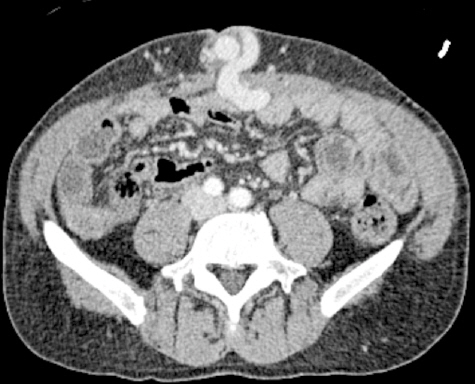
Cross section view of umbilical vein herniating through the umbilical defect.

Sagittal view of umbilical vein herniating through the umbilical defect.
Diagnosis of incarcerated umbilical hernia was subsequently ruled out and no surgical intervention was offered. Our patient instead received medical management with nadolol with clinical resolution of peri-umbilical pain within 3 months.
Discussion
We are reporting a case of an umbilical varix hernia through umbilical defect, with striking similarities to incarcerated bowel hernia. There are two case reports [1,2] with similar findings. However, in our case, herniation of enlarged umbilical vein closely resembles incarcerated bowel through umbilical defect on both physical examination and imaging.
Mainstream treatment of varices secondary to cirrhotic portal hypertension consists of primary prophylaxis of bleeding with either non-selective beta-blocker or endoscopic band ligation and secondary prophylaxis of re-bleeding with combination therapy [3]. In a case of herniated umbilical varix as our patient in this case report; however, we recommend pharmacological management to prevent enlargement of varix and worsening herniation rather than surgical intervention to reduce hernia due to high risk of intra-operative bleeding. Evidence suggests that mortality benefit can be achieved on non-surgical management of varices with hepatic vein pressure gradient (HVPG) reduction of greater than 20% or final HVPG of less than 12 mmHg [4].
Incarcerated umbilical hernia is a surgical emergency. However, in setting of cirrhotic portal hypertension, it is critical to obtain appropriate imaging in order to rule out other differential diagnoses for which surgery is contraindicated, as in our patient.