Retrosternal Hemorrhage after Cardiac Compression
Article information
Body
A 43-year-old male visited the Trauma Center after a pedestrian traffic accident. During initial resuscitation, suddenly the ventricular fibrillation was detected. Cardiac compression and cardioversion were performed for about 10 minutes, and his rhythm was recovered. A chest radiograph was taken (Fig. 1), and focused assessment with sonography in trauma (FAST) was negative (Pericardial effusion wasn’t seen, and heart movement was decreased, However it was difficult to distinguish from the effect of ventricular fibrillation). He was admitted to the intensive care unit and a transesophageal echocardiography (TEE) was performed for cardiac evaluation (Unfortunately, the image wasn’t saved). TEE presented cardiac tamponade features, hence a large amount of fluid was administrated instead of using more inotropic drug. Computed tomography (CT) scan was taken and the CT showed retrosternal hematoma which compressed the right ventricle (Fig. 2). Because retrosternal hematoma was not easily detected in FAST, CT or TEE could be considered for evaluation after cardiac compression [1,2].
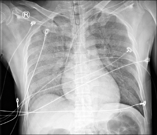
Chest radiograph showed normal cardiac contour.
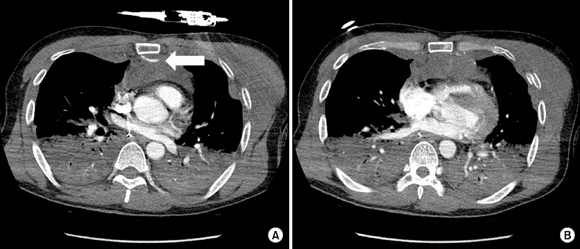
(A) Indicator showed active bleeding of retrosternal area. (B) Heart was compressed by retrosternal hematoma, and both lung atelectasis was shown
Conflicts of Interest
No potential conflict of interest relevant to this article was reported.